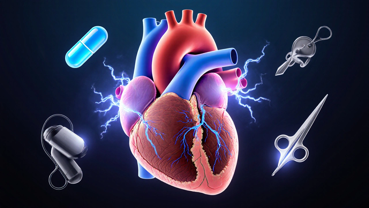
Hypertrophic Subaortic Stenosis is a form of hypertrophic cardiomyopathy in which the interventricular septum thickens enough to narrow the left ventricular outflow tract (LVOT). The blockage forces the heart to work harder and creates an electrical environment that can spark a variety of arrhythmias, from benign premature beats to life‑threatening ventricular tachycardia.
Quick Takeaways
- LVOT obstruction is the main driver of symptoms and arrhythmia risk in HSS.
- Beta‑blockers and calcium‑channel blockers remain first‑line drugs, but myosin inhibitors such as mavacamten are reshaping medical therapy.
- Implantable cardioverter‑defibrillators (ICDs) are the cornerstone for primary and secondary sudden cardiac death prevention.
- Surgical septal myectomy and alcohol septal ablation provide durable relief of obstruction when medication fails.
- Genetic testing and family screening guide risk stratification and long‑term follow‑up.
Understanding the Link Between HSS and Arrhythmias
The thickened septum creates abnormal blood flow patterns and increases wall stress, which in turn alters ion channel behavior. This mechanical‑electrical feedback, often called "mechanotransduction," promotes ectopic foci in the left ventricle and the papillary muscles. Moreover, myocardial fibrosis-visible on cardiac MRI-provides a substrate for re‑entry circuits that can sustain ventricular tachycardia.
Hypertrophic Cardiomyopathy (HCM) is the broader disease class; HSS is the obstructive phenotype. While HCM patients may experience atrial fibrillation due to left‑atrial enlargement, HSS patients are especially prone to ventricular arrhythmias because the outflow‑tract gradient amplifies sympathetic activation during exertion.
Risk Assessment & Stratification
Risk stratification aims to identify who will benefit most from an ICD. The most widely used models incorporate:
- Maximum wall thickness (≥30mm raises risk dramatically).
- History of unexplained syncope.
- Non‑sustained ventricular tachycardia on 24‑hour Holter.
- Abnormal blood pressure response to exercise.
- Family history of sudden cardiac death.
Cardiac MRI quantifies late‑gadolinium enhancement (LGE); LGE >15% of myocardial mass correlates with a two‑fold increase in ventricular arrhythmia incidence. Genetic testing for pathogenic sarcomere mutations (e.g., MYH7 or MYBPC3) refines risk estimates, especially in younger patients.
Pharmacologic Management
Medical therapy focuses on reducing LVOT gradients and stabilizing the cardiac rhythm. The treatment ladder usually follows:
- Beta‑blockers (e.g., propranolol, metoprolol) lower heart rate, prolong diastole, and blunt the gradient.
- Non‑dihydropyridine calcium‑channel blockers (e.g., verapamil) improve diastolic filling and can relieve symptoms.
- Disopyramide, a ClassIa antiarrhythmic, offers both negative inotropy and rhythm control but requires careful monitoring of anticholinergic side effects.
- For refractory ventricular arrhythmias, amiodarone or sotalol may be added, though long‑term toxicity limits use.
- Mavacamten, a selective myosin inhibitor, reduces hypercontractility and has shown significant gradient reduction in the EXPLORER‑HCM trial. It is now FDA‑approved for obstructive HCM and is increasingly used as a bridge to invasive procedures.
Medication choice hinges on symptom severity, blood pressure, comorbidities, and patient preference. Regular echocardiography every 6-12months gauges gradient response and guides dose adjustments.

Device Therapy: When an ICD Becomes Essential
An Implantable Cardioverter‑Defibrillator (ICD) is the only proven intervention to prevent sudden cardiac death in high‑risk HSS patients. Indications include any of the following:
- Prior cardiac arrest or sustained ventricular tachycardia.
- Two or more major risk factors from the stratification list.
- Extensive LGE on MRI (>15% of mass) even without documented arrhythmia.
Modern sub‑cutaneous ICDs avoid trans‑venous leads, reducing infection risk-a notable advantage for young, active patients. Device programming now emphasizes discrimination algorithms that minimize inappropriate shocks, a common quality‑of‑life concern.
Septal Reduction Strategies
When gradients remain >50mmHg despite optimal medical therapy, invasive options are considered. Two main techniques dominate:
| Attribute | Septal Myectomy | Alcohol Septal Ablation |
|---|---|---|
| Invasiveness | Surgical (open‑heart) | Percutaneous catheter‑based |
| Immediate Gradient Reduction | 70-80% | 50-70% |
| Long‑Term Survival | Comparable to general population | Slightly lower; depends on centre experience |
| Complication Rate | 2-4% (conduction block, ventricular septal defect) | 5-8% (need for pacemaker, ventricular arrhythmia) |
| Eligibility | Patients ≥18y, suitable surgical risk | Patients with suitable septal anatomy, high surgical risk |
Myectomy remains the gold standard for younger patients with severe obstruction and concomitant mitral‑valve abnormalities. Alcohol ablation offers a less invasive alternative for older or high‑risk individuals but carries a higher chance of needing a permanent pacemaker.
Lifestyle, Follow‑Up, and Family Planning
Patients should avoid high‑intensity competitive sports that provoke abrupt heart‑rate spikes. Moderate aerobic activity-like brisk walking or light cycling-is usually safe once symptoms are controlled. Hydration, avoiding stimulants (caffeine, decongestants), and regular sleep patterns also reduce arrhythmia triggers.
Follow‑up protocols typically include:
- Clinical review every 6months (symptom check, blood pressure).
- Echocardiogram annually to reassess LVOT gradient and wall thickness.
- Holter monitoring every 1-2years, or sooner if symptoms change.
- Genetic counseling for affected families; first‑degree relatives should undergo screening echocardiogram and, if indicated, genetic testing.
Pregnant women with HSS need multidisciplinary care. Beta‑blockers are generally safe, but teratogenicity concerns limit the use of disopyramide. ICD interrogation before delivery and careful fluid management during labor are essential.
Emerging Therapies & Future Directions
Beyond myosin inhibition, novel agents aim to modify the underlying sarcomere pathology. Gene‑editing approaches using CRISPR‑Cas9 are in pre‑clinical stages, targeting pathogenic MYH7 mutations. Small‑molecule activators of cardiac myosin‑binding protein C are also under investigation, promising to restore normal contractility while preserving diastolic function.
Remote monitoring platforms that stream ECG and device data to cloud‑based analytics are being trialled to catch arrhythmic events earlier, potentially reducing hospital admissions. As artificial‑intelligence algorithms become more adept at interpreting MRI LGE patterns, risk stratification will become more precise, leading to personalized therapy pathways.

Frequently Asked Questions
What symptoms suggest that hypertrophic subaortic stenosis is worsening?
Increasing shortness of breath on exertion, new chest pressure, fainting spells (syncope), and palpitations that interrupt daily activities usually signal a rising LVOT gradient or emerging arrhythmias. An abrupt change should prompt immediate cardiac evaluation.
How does a beta‑blocker help in HSS‑related arrhythmias?
Beta‑blockers slow the heart rate, lengthen diastole, and reduce the force of contraction. This lessens the pressure gradient across the LVOT and dampens the sympathetic surge that often triggers ventricular ectopy.
When is an ICD recommended over medication alone?
An ICD is advised when a patient has survived a cardiac arrest, has sustained ventricular tachycardia, or possesses two or more major risk factors (wall thickness ≥30mm, family sudden death, significant LGE, etc.). The device directly aborts lethal rhythms, something drugs cannot guarantee.
Can alcohol septal ablation replace surgery for all patients?
Not for everyone. Ablation works best when the hypertrophied segment is accessible via coronary arteries and the patient has a higher surgical risk. Younger patients or those with concomitant mitral‑valve disease usually benefit more from surgical myectomy.
Is genetic testing required for every HSS patient?
Guidelines recommend testing for all individuals with a confirmed diagnosis of HCM, as it clarifies prognosis, informs family screening, and may affect therapeutic choices (e.g., eligibility for clinical trials).
What role does mavacamten play in current treatment algorithms?
Mavacamten directly reduces hypercontractility, lowering LVOT gradients without the need for invasive surgery in many patients. It is typically added after beta‑blockers or calcium‑channel blockers fail to control symptoms, and before considering septal reduction procedures.
How often should follow‑up imaging be performed?
An annual echocardiogram is standard for stable patients. If a new symptom emerges or after any change in therapy, imaging should be repeated within 3-6months to assess gradient response and wall‑thickness progression.

Sue Berrymore
September 26, 2025 AT 15:12Wow, the way the septum thickens feels like a heart‑pounding drama unfolding inside each beat! Keep pushing those beta‑blockers and stay consistent with echo checks – your effort really matters. Remember, every step you take toward controlling the gradient is a victory worth celebrating.
Jeffrey Lee
October 1, 2025 AT 06:19Honestly this article overhypes the new drugs – they’re just hype and cost a fortune. No wonder patients get burned.
Ian Parkin
October 5, 2025 AT 21:25It is commendable that the authors have assembled a comprehensive overview of hypertrophic subaortic stenosis and its arrhythmic complications. The delineation of risk stratification criteria, encompassing wall thickness, syncope history, and late‑gadolinium enhancement, reflects current consensus guidelines. Moreover, the emphasis on genetic testing aligns with contemporary precision‑medicine initiatives. The discussion of pharmacologic options, particularly the inclusion of mavacamten, is timely given recent FDA approval. One must also appreciate the balanced presentation of invasive strategies, wherein septal myectomy and alcohol ablation are compared with appropriate nuance. The tabular comparison provides clarity for clinicians navigating procedural choices. The mention of lifestyle modifications, such as avoidance of high‑intensity sports, underscores the holistic management required for these patients. It is also noteworthy that the authors address special populations, including pregnant women, with sensible precautions. While the article excels in breadth, a deeper exploration of long‑term outcomes after myectomy would be valuable. Likewise, further elaboration on the role of sub‑cutaneous ICDs could enhance the device therapy section. The future directions segment, highlighting gene‑editing and AI‑driven risk modeling, demonstrates forward‑thinking. However, readers should be cautioned that many of these emerging therapies remain investigational. The inclusion of remote monitoring platforms signifies an awareness of evolving digital health trends. Overall, the synthesis of pathophysiology, clinical assessment, and therapeutic pathways renders the manuscript a useful reference. It is hoped that clinicians will integrate these insights into practice to improve patient prognosis. In conclusion, the article stands as a solid contribution to the cardiology literature, warranting dissemination among specialists and trainees alike.
Julia Odom
October 10, 2025 AT 12:32The interplay between mechanical obstruction and electrical instability is fascinating, akin to a finely tuned orchestra suddenly missing a conductor. By meticulously titrating beta‑blockers, clinicians can dampen the sympathetic crescendo that fuels ectopic beats. Furthermore, the article’s vivid description of mechanotransduction helps demystify a complex concept for both novices and experts. Continued research into myosin inhibition promises to add a new movement to this therapeutic symphony.
Danielle Knox
October 15, 2025 AT 03:39Oh great, another “breakthrough” pill-just what we needed to complicate the pharmacy shelves.
ruth purizaca
October 19, 2025 AT 18:45The piece covers the basics but fails to delve into nuanced patient experiences.
Shelley Beneteau
October 24, 2025 AT 09:52Building on the enthusiasm expressed earlier, it is worth noting that regular cardiac rehab can further improve functional capacity in HSS patients, complementing pharmacologic therapy. Incorporating structured exercise under professional supervision helps mitigate the risk of abrupt tachycardic spikes that could precipitate arrhythmias.
Sonya Postnikova
October 28, 2025 AT 23:59Hey, I get the frustration, but the new myosin inhibitors really have shown promise in reducing gradients for many folks 😊. They’re not a magic wand, yet they’re a solid addition when traditional meds fall short.
Anna Zawierucha
November 2, 2025 AT 15:05Sure, because adding another drug to the regimen is exactly what every patient dreamed of when they signed up for a simple heart check‑up.
Mary Akerstrom
November 7, 2025 AT 06:12Listen guys the key is regular follow up keep the echo yearly and monitor any new symptoms early the sooner you catch a change the better you can adjust meds or consider an ICD if needed trust the process and stay proactive
Delilah Allen
November 11, 2025 AT 21:19Indeed-one might argue that the very notion of “monitoring” is a metaphysical assertion of control over the chaotic nature of cardiac electrophysiology; yet without such obsessive vigilance, the heart’s erratic impulses would reign unchecked!; It is in this relentless pursuit of order that we confront the primal disorder within us all!!!
Nancy Lee Bush
November 16, 2025 AT 12:25Absolutely! Frequent check‑ups are the backbone of safe management 😊; they also empower patients by providing tangible data on their progress!!!
Dan Worona
November 21, 2025 AT 03:32What they don’t tell you is that the push for expensive myosin inhibitors is backed by a hidden network of pharma lobbyists with ties to the very insurers who profit from long‑term device implantation. Stay alert, question the hype, and demand transparency.
Chuck Bradshaw
November 25, 2025 AT 18:39In case anyone missed it, the ESC guidelines actually prioritize ICD implantation over septal ablation in patients with ≥2 risk factors, and the cutoff for wall thickness is 30 mm, not 25 mm as some sources mistakenly claim.